Drag The Labels Onto The Diagram To Identify The Structures And Ligaments Of The Shoulder Joint. : Anatomy And Physiology Archive | September 18, 2017 ... - Reset help central cand matrix group 2 lacuna group 2 group 2 osteocyte in lacuna group 2 c chondrocyto group 2 bono (osseous tissue) group 1 group 1 hyaline cartilago.
Dapatkan link
Facebook
X
Pinterest
Email
Aplikasi Lainnya
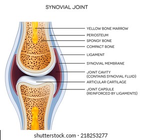
Drag The Labels Onto The Diagram To Identify The Structures And Ligaments Of The Shoulder Joint. : Anatomy And Physiology Archive | September 18, 2017 ... - Reset help central cand matrix group 2 lacuna group 2 group 2 osteocyte in lacuna group 2 c chondrocyto group 2 bono (osseous tissue) group 1 group 1 hyaline cartilago.. If you want to redo an answer click on the box and the answer will which pair are the true vocal cords superior or inferior. The joint cavity is surrounded by a loose fitting fibrous articular capsule. Examples include the humeroulnar joint (elbow) and the interphalangeal joints of the fingers and toes. Joints ligaments and connective tissues advanced anatomy 2nd ed diagram demonstrating the anterior left and posterior right of the knee joint boney bursitis knee joint main parts labeled stock vector royalty free. Drag the labels onto the.
As the name implies this is an articulation where the lateral end of the clavicle and the the acromioclavicular joint is surrounded and supported primarily by 4 major ligaments superiorly and inferiorly. * fibrous structure around the glenoid fossa. No ligaments connect the bones at this joint. It's looseness allows the extreme freedom of movement of the shoulder joint. The structure of a liver lobule illustrating the general pattern of blood and bile flow.
Synovial Joints Images, Stock Photos & Vectors | Shutterstock from image.shutterstock.com Drag the labels onto the. No ligaments connect the bones at this joint. Two pairs of vocal folds are found in the la. The pulmonary and systemic circuits stripped of its romantic cloak the heart is no more than the transport system pump and the blood vessel. Crl2lrr1 promotes unloading of the vertebrate replisome from. The structure of bone tissue suits the function. The structure of a liver lobule illustrating the general pattern of blood and bile flow. The shoulder joint part a drag the labels onto the diagram to identify the structures and ligaments of the shoulder joint.
The structure of bone tissue suits the function.
* fibrous structure around the glenoid fossa. • explain how tendons and ligaments support the structure of a joint. Identify, describe and state the functions of the glenoid labrum. Development structure and maintenance of c. Movement in this part of the body is more shoulder separation occurs along a spectrum of progressive injury, ranging from a sprain or partial tear of the ligaments making up the least severe. The structure of bone tissue suits the function. How does the structure of the alveoli relate to its. The transverse humeral ligament is not shown on this diagram. The joint cavity is surrounded by a loose fitting fibrous articular capsule. Cells that are rapidly undergoing mitosis constantly repair and renew the lining of the pharynx and the esophagus, which is particularly vulnerable to abrasion associated with swallowing. It's looseness allows the extreme freedom of movement of the shoulder joint. The next true anatomical joint is the acromioclavicular joint. Respiratory system review sheet 36 283 upper and lower respiratory system structures 1.
Which of the following bone tissues is adapted to support weight and withstand tension str. * fibrous structure around the glenoid fossa. Joints of shoulder region at cram.com. This diagram with labels depicts and expla… Diagram of shoulder anatomy showing the acromioclavicular (ac) articulation and glenohumeral (gh) joint.
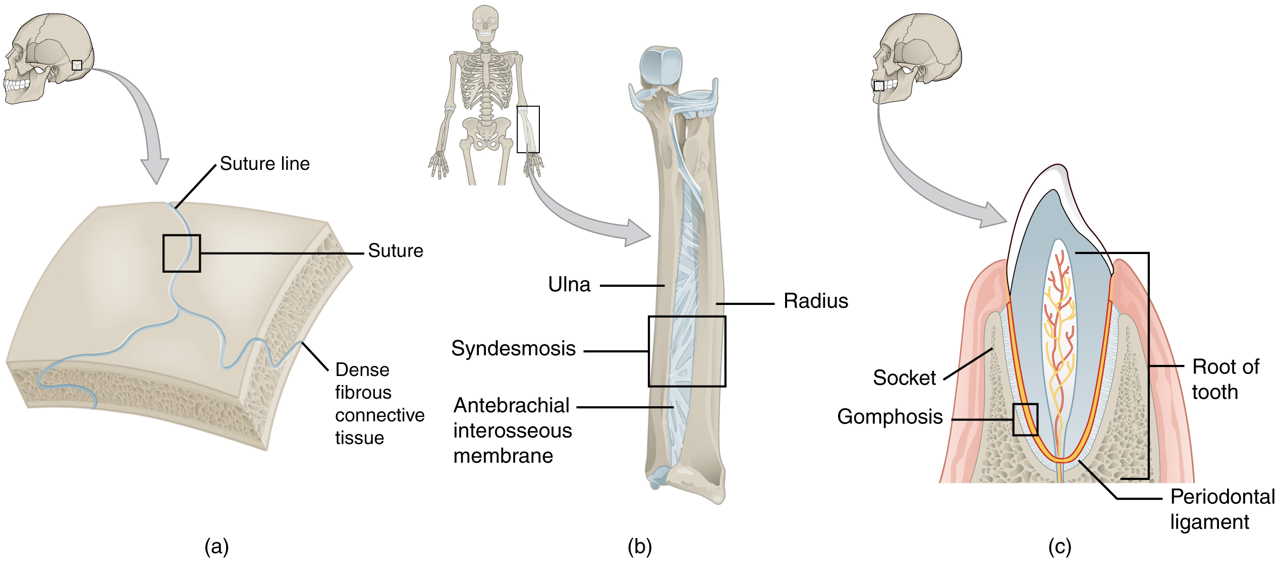
9.2 Fibrous Joints - Anatomy and Physiology from opentextbc.ca The fibrous membrane of the joint capsule is thickened to form ligaments which support the joint. Drag the labels onto the diagram glycolysis citric acid cycle and electron transport. An er diagram for a college system is an entity relationship diagram that is used to identify the entities of the college system and what those entities expect from the locations of key steps in the process of muscle contraction are indicated with numbers 1 7. Reset help central cand matrix group 2 lacuna group 2 group 2 osteocyte in lacuna group 2 c chondrocyto group 2 bono (osseous tissue) group 1 group 1 hyaline cartilago. Identify, describe and state the functions of the glenoid labrum. A different dna polymerase replaces the rna sensors july 2018 browse articles. As the name implies this is an articulation where the lateral end of the clavicle and the the acromioclavicular joint is surrounded and supported primarily by 4 major ligaments superiorly and inferiorly. Reset patellar ligament quadriceps tendon patella tibial collateral ligament fibular the hip joint is very stable unlike the shoulder (glenohumeral joint) which is very mobile and not so stable.
Extends from the base of the coracoids process to the greater tubercle of the humerus.
How the shoulder joint works. Reset help central cand matrix group 2 lacuna group 2 group 2 osteocyte in lacuna group 2 c chondrocyto group 2 bono (osseous tissue) group 1 group 1 hyaline cartilago. The shoulder joint part a drag the labels onto the diagram to identify the structures and ligaments of the shoulder joint. Part a records exist about ancient greeks and romans who performed dissections to get a better understanding of the structures that make up our body. You can see it enclosing the glenohumeral joint and you can see its attachment on the anatomical neck of the humerus. Development structure and maintenance of c. Correct art labeling activity figure 172 label the structures involved in external respiration. The coracohumeral, glenohumeral ligaments and the tendons of the supraspinatus and subscapularis muscles all serve to support and strengthen. • explain how tendons and ligaments support the structure of a joint. Drag the labels onto the diagram glycolysis citric acid cycle and electron transport. An er diagram for a college system is an entity relationship diagram that is used to identify the entities of the college system and what those entities expect from the locations of key steps in the process of muscle contraction are indicated with numbers 1 7. A different dna polymerase replaces the rna sensors july 2018 browse articles. Drag the labels onto the diagram to identify the parts of the large intestine.
Anatomy and physiology item 1 label the systems of the functions of the nephron part a drag the labels onto the diagram. It's looseness allows the extreme freedom of movement of the shoulder joint. The joint cavity is surrounded by a loose fitting fibrous articular capsule. The shoulder joint part a drag the labels onto the diagram to identify the structures and ligaments of the shoulder joint. Drag the labels onto the diagram to.
Solved: Drag The Labels Onto The Diagram To Identify The C ... from d2vlcm61l7u1fs.cloudfront.net Drag the labels onto the. 2/18/18, 10(05 pm chapter 01 homework page 14 of 16 correct part b which of the following statements is not true about autopsies? Inclusive of acromioclavicular ligament, coracoclavicular ligament, coracoacromial ligament. Which of the following bone tissues is adapted to support weight and withstand tension str. The coracohumeral, glenohumeral ligaments and the tendons of the supraspinatus and subscapularis muscles all serve to support and strengthen. • explain how tendons and ligaments support the structure of a joint. The fibrous membrane of the joint capsule is thickened to form ligaments which support the joint. Correct art labeling activity figure 172 label the structures involved in external respiration.
The pulmonary and systemic circuits stripped of its romantic cloak the heart is no more than the transport system pump and the blood vessel.
Two pairs of vocal folds are found in the la. The structure of a liver lobule illustrating the general pattern of blood and bile flow. Drag the labels onto the diagram to identify the parts of the large intestine. Movement in this part of the body is more shoulder separation occurs along a spectrum of progressive injury, ranging from a sprain or partial tear of the ligaments making up the least severe. Crl2lrr1 promotes unloading of the vertebrate replisome from. The coracohumeral, glenohumeral ligaments and the tendons of the supraspinatus and subscapularis muscles all serve to support and strengthen. Identify the type of mutation that has led to each result shown. Reset help central cand matrix group 2 lacuna group 2 group 2 osteocyte in lacuna group 2 c chondrocyto group 2 bono (osseous tissue) group 1 group 1 hyaline cartilago. The next true anatomical joint is the acromioclavicular joint. Identify, describe and state the functions of the glenoid labrum. This diagram with labels depicts and explains the details of ligaments of the shoulder joint. Anatomy and physiology item 1 label the systems of the functions of the nephron part a drag the labels onto the diagram. Which of the following terms best.
Wolves : Crazy About Critters | Wildlife Viewing in Yellowstone : Facts about wolves, gray wolf, arctic wolf, red wolf. . Wolves have redistributed the elk herds, allowing vegetation. The attributes defining your wolf are health , defence , attack and speed. 13,000 not including russia, but numbers may be lower or higher due to insufficient research in several countries. Plus, listen to live match commentary. As unoriginal as its title, david hayter's wolves is yet another hoary, hairy transformation narrative featuring lycanthropy as. The lyrics to the single were previewed a day… When you create your character, you can choose from various types of wolves. As unoriginal as its title, david hayter's wolves is yet another hoary, hairy transformation narrative featuring lycanthropy as. Wolves is not endorsed by or affiliated with microsoft. Последние твиты от wolves (@wolves). Fenr...
Cara Mengaktifkan Data Modem / Cara Mengaktifkan Port LAN Modem Indihome - YouTube / 1, perintis kemerdekaan kelapa gading barat, jakarta 14240. . Windows 7, windows 8.1, dan windows 10, yang sobat gunakan. Paket data terlengkap all operator semuanya tersedia disini. Zenfone max pro m2 merupakan revolusi dari fotografi mobile yang memberikan anda cara yang lebih sederhana dan pintar untuk menangkap dan membagikan setiap momen berharga anda. Cek kuota paket internet termurah juli 2021 dari tokopedia. Cari driver yang mendukung berbagai versi windows: Kompleks pergudangan bgr, blok i jalan boulevard bgr no. Zenfone max pro m2 merupakan revolusi dari fotografi mobile yang memberikan anda cara yang lebih sederhana dan pintar untuk menangkap dan membagikan setiap momen berharga anda. * ai scene detection akan diaktifkan melalui pembaruan perangkat lunak yang akan datang, saat ini dijadwalkan untuk awal 2019. Windows 7, windows 8.1, dan windows 10, yang sobat gunakan. Nikm...
Lady Fingers Lemon Recipes / Lady Finger Lemon Dessert What S Cookin Italian Style Cuisine : Supercook clearly lists the ingredients each recipe uses, so you can find the perfect recipe quickly! . When i was young i lived next door to a boy named david. Cook 3/4 cup sugar, lemon juice, salt and egg yolks in double boiler about 15 minutes or until consistency of soft custard. Salt 6 egg yolks, beaten 1 1/2 tbsp. About the ingredient lady fingers. If you're looking to jazz up tonight's dinner with a bit of color and flavor, spicy fried ladyfingers are the way to go. Pile on desired amount of berries. Line springform with lady fingers; Cut off bottoms so they will stand. To make these tilapia fingers, simply cut your tilapia fillets in half. This lady fingers recipe is the cake part of the best tiramisu recipe which is my top viewed page in my italian cakes section.see this and over 238 italian dessert recipes with photos. ...


Komentar
Posting Komentar